Nowadays, more and more neurological disorders can be detected more easily and at an earlier stage thanks to the ultra-performant devices. One of the most used devices in neurology is the electromyograph (EMG).
The investigation with the EMG is called electromyography; it records the electrical activity of nerves and muscles. When they are active, the muscles produce electrical impulses directly proportionally to the level of muscle contraction.
Specialists use this test to detect abnormal electrical activities in the muscles caused by diseases such as muscular inflammation, muscular dystrophy, amyotrophic lateral sclerosis, peripheral nerve damage, herniated disc, etc. Photo source: https://www.nni.com.sg
When an EMG electromyography is recommended
There are many neurological disorders that can be diagnosed by an electromyography, and here are some cases where it is essential:
- establishing unexplained causes of muscle weakness;
- determining the differences between muscle weaknesses caused by peripheral muscle disorders and those caused by nervous system disorders;
- establishing the differences between muscle weakness caused by the actual diseases and those caused by the muscles not functioning at normal parameters;
- differentiation between a psychosomatic disorder, a central nervous system (encephalus and spinal cord) disorder, or a peripheral neurogenic syndrome.
What is the EMG test?
An electromyography is performed in two stages:
The first stage of the investigation is the evaluation of nerve conduction on sensory and motor nerves. For this, some electrodes are used as small disks that stick to the surface of the skin. They record the potential problems of the electrically stimulated nerves. The computer calculates the speed of nervous impulses, but also other functional parameters of the nerve activity.
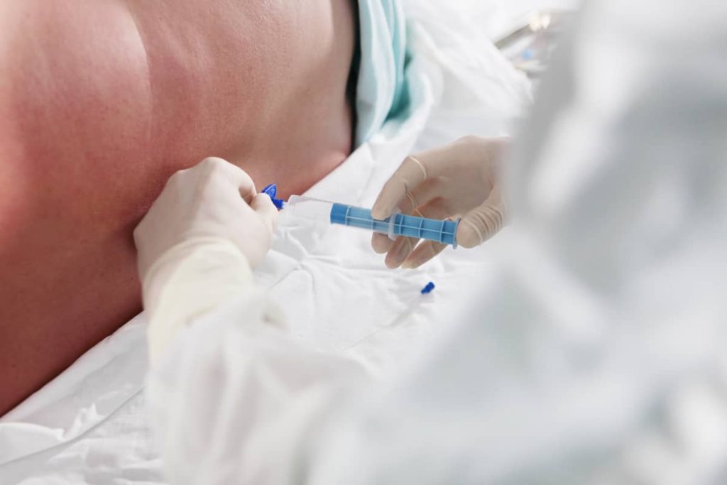
The second stage of the investigation consists of the insertion of a needle (disposable) with electrode function in the muscle mass. The procedure can cause mild or moderate pain, as well as a slight bleeding. In patients undergoing anticoagulant therapy, this investigation is contraindicated. In such cases, it is recommended, at the advice of a specialist, to discontinue the treatment with anticoagulants about 4-5 days before the investigation. The electrical signals that the electrode needle records are analysed by a computer that provides important information about nerves and muscles.
Risks
The EMG electromyography test presents minor risks. In most cases, patients experience mild muscle pain over a few days.
Types of investigations with EMG electromyography
Several types of investigations can be performed with this device. Depending on the symptoms and their history, one can choose the following: VCS / VCM needleless electroneurography (ENG), EMG needle electromyography, repetitive electric stimulation electromyography, tremor analysis electromyography and electromyography with vegetative tests.